【所属科室】
消化科
【基本资料】
患者,男,61岁
【主诉】
体检时发现便中带血1月余。
【现病史】
患者1月前因体检时查便常规发现便中带血,遂至外院行肠镜检查,病理结果提示:直肠癌。
【影像图片】

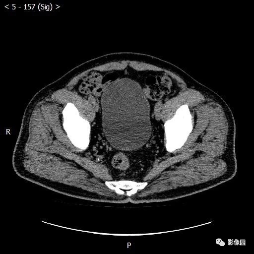
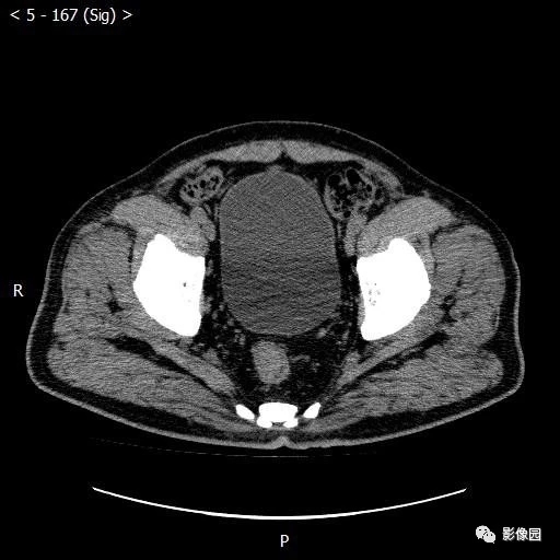

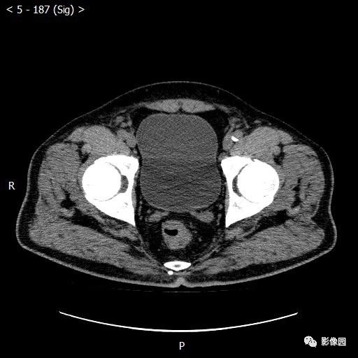
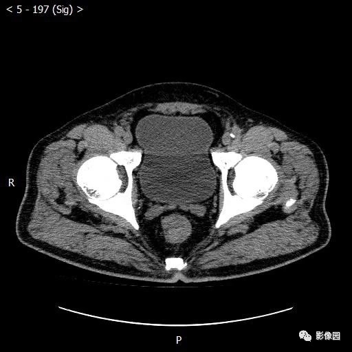
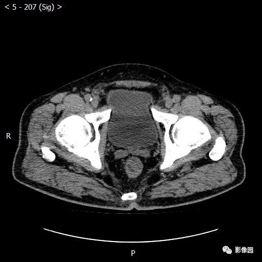
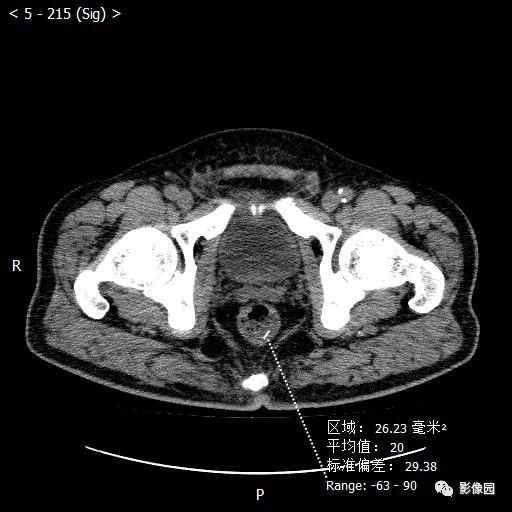
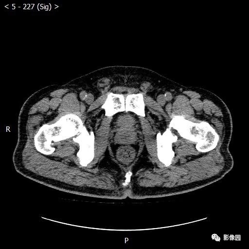


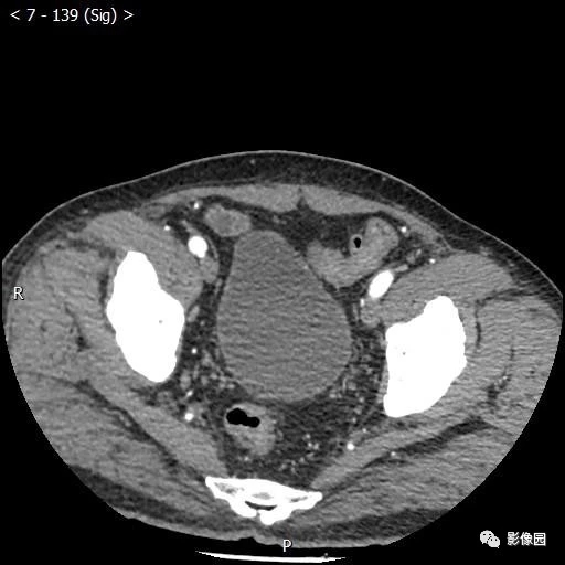

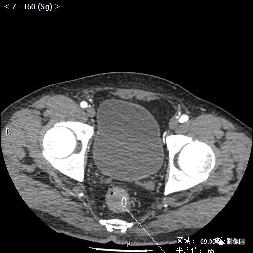
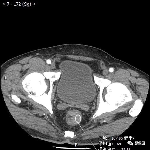
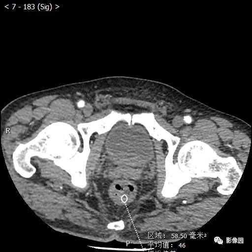
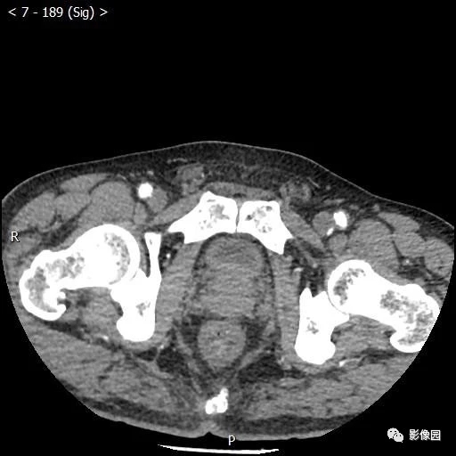
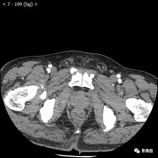

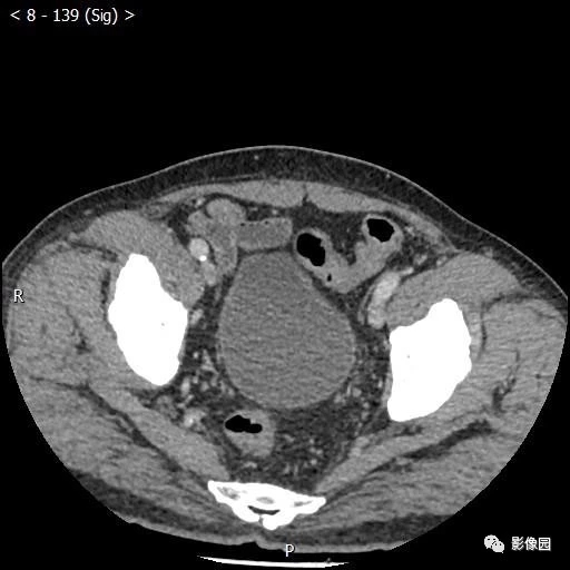
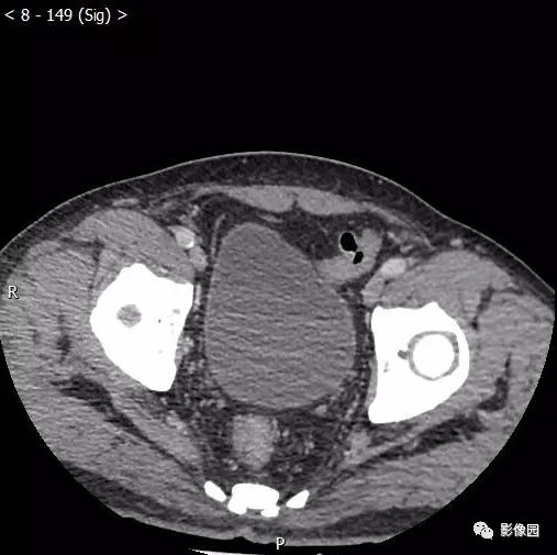

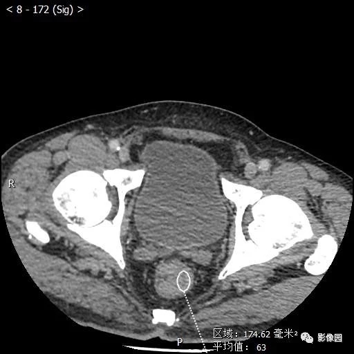
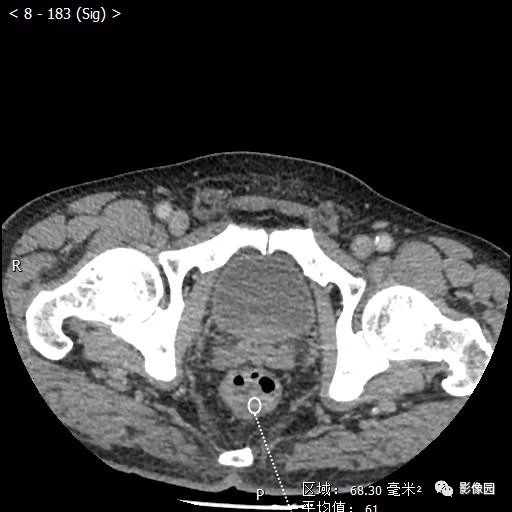
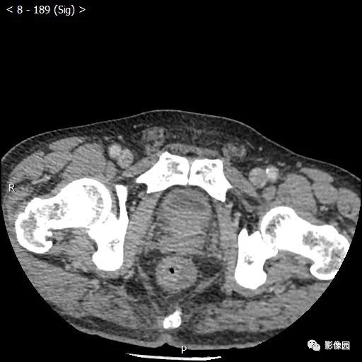
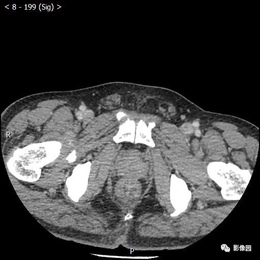
【讨论】
评论:直肠壁不均匀增厚,以左侧为著,部分突向肠腔,部分浆膜层周围脂肪间隙模糊,考虑恶性病变。
【结果】
病理诊断:中分化腺癌,部分为黏液腺癌,癌浸润肠壁全层,未侵及周围脂肪组织,未见脉管癌栓及神经侵犯,肠周淋巴结未见癌转移(0/22)。
【病例小结】
大肠癌包括结肠癌(carcinoma of colon)及直肠癌(colorectal carcinoma),发生部位约半数以上位于直肠,20%位于乙状结肠,其次依次为盲肠、升结肠、降结肠、横结肠。发病年龄多在40~60岁,但30岁以下的青年大肠癌并不少见。临床表现:早期常表现为粪便隐血阳性;随后可出现排便习惯及粪便性状改变;腹痛;局部肿块及全身情况改变(右半结肠癌多见)。临床上习惯用Dukes分期:A期:癌局限于肠壁;B期:癌穿透浆膜;C期:有局部淋巴结转移;D期:有远处转移。
影像学诊断要点:结肠镜检及活检有助于确诊。X线:增生型:主要表现为充盈缺损,充盈缺损周边的的粘膜破坏中断或见小溃疡。气钡双重造影可显示肿块的轮廓。溃疡型:主要表现为腔内突起的龛影和“半月征”。浸润型:主要沿肠壁环形生长,使肠壁增厚,肠腔狭窄,可见狭窄段粘膜呈锯齿状。混合型:常有两种以上的表现混合存在。
CT表现:
(1)肠壁增厚,增厚的肠壁黏膜面多明显凹凸不平。
(2)腔内肿块影,偏心性,呈分叶状或不规则形,与正常肠壁分界清楚,肿块表面可见小溃疡,呈火山口样。
(3)肠腔狭窄,且为非对称性。
(4)增强扫描可见较明显异常强化。
(5)浆膜及临近器官受侵表现。
MRI表现:
T1WI低或等软组织信号影,T2WI肿瘤信号增高,接近或高于脂肪组织的信号强度,增强扫描可见轻-中度强化。其他表现同CT。
鉴别诊断:
1、与慢性结肠炎相鉴别:后者狭窄段肠壁较光滑,形态可变,无肿块影。
2、与淋巴瘤相鉴别:后者常发生于回盲部,CT上可见局部肿块和肠壁增厚,轮廓较光整,少有毛刺及周围浸润表现,常伴腹腔、盆腔及腹膜后淋巴结肿大,并可融合成团。淋巴瘤病程长,病变广泛,但无肠梗阻表现为本病特点。
3、与结肠良性肿瘤或息肉:后者的充盈缺损表现光滑整齐,粘膜规则,蠕动正常,而前者多表现为粘膜皱襞破坏中断,管壁僵硬。
4、与肠结核相鉴别:后者常累及末端回肠与盲肠,盲肠缩短挛缩,一般不见充盈缺损征象。多有肠外结核病史,PPD试验阳性有助于确诊。
作者:佚名
版权声明:
本网站所有注明“来源:梅斯医学”或“来源:MedSci原创”的文字、图片和音视频资料,版权均属于梅斯医学所有。非经授权,任何媒体、网站或个人不得转载,授权转载时须注明“来源:梅斯医学”。其它来源的文章系转载文章,本网所有转载文章系出于传递更多信息之目的,转载内容不代表本站立场。不希望被转载的媒体或个人可与我们联系,我们将立即进行删除处理。
在此留言


#老年男性#
39